Home
Uncategories
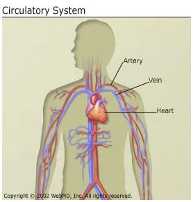
Arteries Diagram Easy / Human Circulatory System - Organs, Diagram and Its Functions / These vessels are channels that distribute blood to the body.
Arteries Diagram Easy / Human Circulatory System - Organs, Diagram and Its Functions / These vessels are channels that distribute blood to the body.
Arteries Diagram Easy / Human Circulatory System - Organs, Diagram and Its Functions / These vessels are channels that distribute blood to the body.. This makes it more difficult for blood and oxygen to get through the arteries to the body's important organs. This is known as the main pulmonary artery or pulmonary trunk. The narrowed arteries are at higher risk for complete blockage from a sudden. Supply oxygenated blood to the head and neck regions of the body. The aorta is the main systemic artery and the largest artery of the body.
See the back for a diagram showing the two circulation routes. It is returned to the heart in the veins. Arteriosclerosis occurs when the blood vessels that carry oxygen and nutrients from your heart to the rest of your body (arteries) become thick and stiff — sometimes restricting blood flow to your organs and tissues. Smartdraw has a number of templates included for circulatory system diagrams, cardiovascular system diagrams, blood circulation diagrams, and more. Original vintage human anatomy victorian bookplate print 1890s medical diagram veins arteries blood circulatory system of the human body thepapermuseum.
Simple Heart Diagram label | Heart diagram, Human heart diagram, Simple heart diagram from i.pinimg.com This area is known as the circle of willis. Arteries and veins are two of the body's main type of blood vessels. Arteries carry blood away from the heart in two distinct pathways: Comprised of the heart, blood vessels and the blood itself, it is divided into two loops which. A very good labled with diagram easy to study. An easy way to jog. It is returned to the heart in the veins. The name pulmonary artery is easy to remember because it carries blood to the lungs, so that will help you think of pulmonary.
Human anatomy for muscle, reproductive, and skeleton.
It is a central communication that unites the internal carotid and vertebrobasilar systems. Arteries and veins are two of the body's main type of blood vessels. Arteries of the brain and 'circle of willis' diagram there is a point at which the anterior and posterior arterial circuits of the brain unite or anastomose. See more ideas about arteries anatomy, arteries, echocardiogram. When a person has arteriosclerosis , the walls of their arteries get harder, stiffer, and less stretchy. Small branches dive into the heart muscle to. This area is known as the circle of willis. These vessels are channels that distribute blood to the body. Arteries are blood vessels that carry blood away from the heart to other tissues. This is known as the main pulmonary artery or pulmonary trunk. The narrowed arteries are at higher risk for complete blockage from a sudden. Molly smith dipcnm, mbant • reviewer: When the coronary arteries narrow to the point that blood flow to the heart muscle is limited (coronary artery disease), collateral vessels may enlarge and become active.
Coronary arteries supply blood to the heart muscle. Cardiovascular system diagrams, quizzes and free worksheets. These vessels are channels that distribute blood to the body. This is known as the main pulmonary artery or pulmonary trunk. A very good labled with diagram easy to study.
Heart - coronary arteries Illustrations from site.motifolio.com Amicus illustration of amicus,anatomy,brain,arterial,artery,arteries,supply,cerebral,cavernous the right and left upper and lower limbs create a flow chart showing the major systemic veins through which blood travels… This is done to make it easy to distinguish between arteries and veins as they look almost identical. Arteriosclerosis occurs when the blood vessels that carry oxygen and nutrients from your heart to the rest of your body (arteries) become thick and stiff — sometimes restricting blood flow to your organs and tissues. All the required arteries and veins on the pancake man. A very good diagram, made the study of coronary arteries of anterior heart wall very easy. Artery, in human physiology, any of the vessels that, with one exception, carry oxygenated blood and nourishment from the heart to the tissues of the body. Heart anatomy, video, quiz, and chart included! An easy way to jog.
Cardiovascular system diagrams, quizzes and free worksheets.
An easy way to jog. The capillaries connect the two types of blood. Diagram of blood vessel types each time your heart beats, blood is forced into large arteries. It originates from the heart and branches out into smaller arteries which supply blood to the head region (brachiocephalic artery), the heart itself (coronary arteries), and the lower regions of the body. Cardiovascular system diagrams, quizzes and free worksheets. Dimitrios mytilinaios md, phd last reviewed: Blood supply to the scalp. Small branches dive into the heart muscle to. When a person has arteriosclerosis , the walls of their arteries get harder, stiffer, and less stretchy. May be an important source of collaterals. For more anatomy content please follow us and visit our website: 5 minutes the cardiovascular system is a vital organ system which is quite literally at the centre of everything. A very good diagram, made the study of coronary arteries of anterior heart wall very easy.
This is done to make it easy to distinguish between arteries and veins as they look almost identical. Carries deoxygenated blood from the right ventricle to the lungs. Molly smith dipcnm, mbant • reviewer: For more anatomy content please follow us and visit our website: Human anatomy for muscle, reproductive, and skeleton.
Anatomy and Circulation of the Heart from img.webmd.com A very good diagram, made the study of coronary arteries of anterior heart wall very easy. This makes it more difficult for blood and oxygen to get through the arteries to the body's important organs. These vessels are channels that distribute blood to the body. Blood supply to the scalp. Great for usmle, nursing, students, doctors, and medical learners. Carries deoxygenated blood from the right ventricle to the lungs. Molly smith dipcnm, mbant • reviewer: When the coronary arteries narrow to the point that blood flow to the heart muscle is limited (coronary artery disease), collateral vessels may enlarge and become active.
Blood flow through the heart made easy with a simple diagram of the cardiac circulation pathway and steps in order.
Great for usmle, nursing, students, doctors, and medical learners. Artery, in human physiology, any of the vessels that, with one exception, carry oxygenated blood and nourishment from the heart to the tissues of the body. Supply oxygenated blood to the head and neck regions of the body. A very good labled with diagram easy to study. Arteries carry blood away from the heart in two distinct pathways: How to make a circulatory system diagram. Arteriosclerosis occurs when the blood vessels that carry oxygen and nutrients from your heart to the rest of your body (arteries) become thick and stiff — sometimes restricting blood flow to your organs and tissues. Arteries and veins are two of the body's main type of blood vessels. Learn the differences between an artery and a vein. See more ideas about arteries, anatomy and physiology, medical anatomy. See more ideas about arteries anatomy, arteries, echocardiogram. Blood is transported in arteries, veins and capillaries. Original vintage human anatomy victorian bookplate print 1890s medical diagram veins arteries blood circulatory system of the human body thepapermuseum.
Carry oxygenated blood from the abdominal aorta to the legs and feet arteries diagram. Blood supply to the scalp.


0 Comments:
Post a Comment